

Molecular and structural biologists from Nanyang Technological University, Singapore (NTU) have discovered a new way for disabling respiratory syncytial virus (RSV) and human metapneumovirus (HMPV) after shedding light on a key part in the structure of the viruses.
RSV and HMPV are similar viruses that can lead to highly dangerous respiratory diseases such as pneumonia and bronchiolitis found in the following groups of people: premature babies and infants, elderly, and people with a weak immune system.
When these viruses infect the human cells, they take control over the cell’s machinery and duplicate themselves. This infection process is started when special proteins released by the virus communicate with one another to make individual protein complexes.
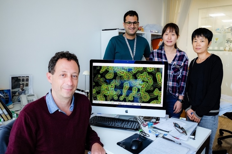
The research team from NTU used cryo-electron microscopy to create the image of the molecular structure of one of the large complexes which is an enzyme called HMPV L:P polymerase.
This method uses an advanced electron-scanning microscope, which can project a cryogenically frozen sample down to the sub-nanometre range, which is about ten times smaller than a strand of human DNA or one million times smaller than the width of a human hair.
The images of the HMPV L:P polymerase enzyme were produced at a resolution of 3.7 Angstrom (0.37 nanometres) and as two-dimensional pictures.
Based on these images, the team then created three-dimensional computer models of the proteins’ L:P molecular structures.
Analysis done of these structures showed results of the key locations for the molecules to communicate with one another. This provided insights into the new targets for developing antiviral molecules against both viruses.
Dr Lescar, Principal Investigator at the NTU Institute of Structural Biology, shared that this information is crucial to researchers for developing inhibitors that interrupt the enzymatic activities of HPMV L:P protein and possibly block infection by the virus.
He hopes that this information will be beneficial to researchers in pharma and academia from around the world to design important therapies for difficult viral infections that often result in antibiotic-resistant bacterial infections.
They hope that that the inhibitors can also be used against a variety of viruses from respiratory diseases and for other viral diseases too.
NTU’s scientists have previously made such strides for discoveries in the field of health technology.
In an earlier article. OpenGov reported that Scientists from the Nanyang Technological University of Singapore (NTU Singapore) have created a lab-on-a-chip system that can detect the health features of an individual’s immune system. This can be done within minutes with just a drop of their blood.
The system employs a combination of microfluidics, which are tiny microscopic channels that can isolate white blood cells from blood, and electrical sensors to create the chips.
These chips will be able to identify the variations in the electrical properties of white blood cells which are collected from both healthy and diabetic patients.
Upon further successful laboratory and clinical assessments, this system could potentially be turned into a portable device for use in family clinics and polyclinics.
The device could in the future aid doctors to quickly obtain insights into a person’s immune system and detect early signs of inflammation and infection that could indicate the need for more in-depth examinations of an individual.
Immune health is a sign of cardiovascular diseases. Scientists can potentially use the device as an additional screening tool for the early detection of heart diseases. Cardiovascular diseases were the cause of about 30.1 percent of all deaths in 2017 in Singapore.