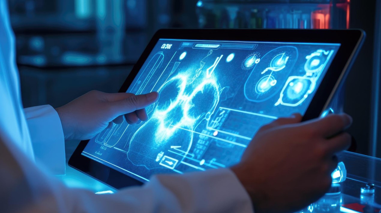


Researchers at the Auckland Bioengineering Institute (ABI) and the Department of Engineering Science are combining machine learning and state-of-the-art imaging to develop an automated analysis technique that will radically improve the diagnosis and treatment of breast cancer.
As reported, researchers from New Zealand’s University of Auckland have received NZ$ 1.05 million in philanthropic funding to advance research, in which they have developed biomechanical analysis techniques that automatically merges information from different medical images of the breast.
Benefits
This will provide clinicians with more information about any abnormality, as suspicious lesions and warning signs for cancer can appear differently in the various types of images.
An example of how it can help is by co-locating abnormalities like micro-calcifications with regions of increased blood supply identified using MRI and which can also indicate tumour growth.
Micro-calcifications are tiny and hard-to-detect features visible on x-ray mammograms indicative of early stages breast cancer.
The Problem
One of the key issues with merging information from current medical imaging technologies is that they scan the breast in different positions.
In an X-ray mammogram, which is the gold standard for the early detection of breast cancer, a patient is standing and the breast is compressed between two plates.
If she is referred for ultrasound imaging, the image is taken while the patient lies on her back. In magnetic resonance imaging (MRI), the images are taken typically with the patient lying on her stomach.
As the breast is a soft organ that changes shape according to the position in which it is held, cancerous lesions can move around in response to the external loads.
This can make it difficult for clinicians, surgeons and radiologists to identify the exact region that needs further investigation or treatment.
After a mammogram, follow-up imaging known as ‘second look ultrasound’ can involve clinicians spending up to an hour, just trying to find, with some degree of certainty, the same piece of abnormal tissue that they spotted on a mammogram.
The Solution
The use of biomechanics will predict the movements of those suspicious tissues. Thus, when a person is lying on her back, such as during surgery, the surgeon can be provided with a much better idea as to where to find the suspicious lesion.
Part of the challenge has been identifying the different biomechanical properties of the different types of breast tissues in order to account for the individuality of each patient.
Breasts come in all shapes and sizes and the mechanical properties of the tissues vary from person to person. The challenge is to create realistically, individual specific models.
The team has been able to draw on 200 scans, with patient permission, provided by the Auckland District Health Board.
The success of their research can be attributed to a long collaboration with an MRI expert at Auckland City Hospital.
The expert shared that the researchers have made enormous progress in developing methods for analysis and mathematical modelling of breast tissue.
It has been a long-standing, close, and mutually beneficial collaboration, in which clinicians are learning more about mathematical modelling, and the bioengineering researchers are learning more about the real world challenges facing clinicians working with breast cancer.