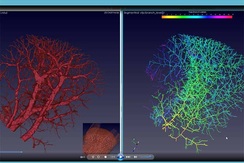
A comparison of the enhanced blood vessel prior to skeletonisation (on the left) and after the end-point skeletonisation process (on the right) (Image credit: CSIRO)
Researchers from Data61 in Australia, in collaboration with researchers at the Shanghai Institute of Applied Physics, Chinese Academy of Sciences, have developed an algorithm which could improve the detection of angiogenesis, the development of new blood vessels which is known to precede the growth of cancers. Angiogenesis plays a critical role in the growth and spread of cancer. A blood supply is necessary for tumors to grow beyond a few millimeters in size and spread through the body.
Until now, images of blood vessel structure taken from high-resolution imaging have only been able to produce a skeletonised view of blood vessel structure which provided limited detail and accuracy. The algorithm can generate an accurate representation of vasculature (blood vessels), while preserving the length and shape information of the blood vessel and its branches.
This development was done using a technique called end-point constraints. End-points are critical in preserving the geometrical features of new blood vessels, including branching patterns and the lengths of terminal vessels.
The accurate quantification of vasculature changes, particularly the number of terminal vessel branches, can play a critical role in accurate assessment and treatment of cancers. Earlier detection of blood vessel growth may therefore lead to a faster diagnosis of malignant tumour growth, which is a key factor in successful treatment and patient survival. During anti-angiogenesis treatment also, continuously monitoring subtle proliferations in blood vessels over time is crucial, because patients may react to the treatment differently.
During the study the researchers produced images of the brains and livers of mice at various stages of cancer growth.They analysed 26 high-resolution 3D micro-CT images from 26 mice, produced by the Shanghai Synchrotron Radiation Facility (SSRF) and using the images developed the algorithm for representation of the blood vessels.
The new software, allows researchers to measure subtle changes in the proliferation of blood vessels, including the number and length of the blood vessel branches, and produces significantly clearer skeletons of the vasculature than previously possible.
Cancer Council Australia CEO Professor Sanchia Aranda said, "This exciting project seeks to bring to life the tumour micro-environment through 3D synchrotron images of the vessels and will help to advance our understanding of this critical cancer progression process."
Next steps
The Shanghai Synchrotron Beamline used to produce the images generates radiation levels unsafe for human imaging. In order to conduct clinical trials in humans, the researchers are looking for 3D imaging technologies and partnering with a hardware manufacturer that can produce high-resolution images with safe levels of radiation for humans.
Dr. Dadong Wang, lead researcher on this project from Data61’s Quantitative Imaging team, said that the applications of the software could be extended beyond 3D angiogenesis analysis, in areas such as drug development.
Dr. Wang said, “While there is great interest in taking these findings further, there is still a long way to go before this new development can be applied to human patients. But we are very hopeful, and currently looking for collaborators and partners to take the technology to the next stage."


